Français
 Deutsch
 Español
 Italiano
 Nederlands
 Polski
 Русский
 Svenska
 Português
 Hrvatski
 Ελληνικα
 简体中文
简体中文 العربية
ECG and heart rhythm monitoring
Electrocardiogram (ECG)
The most important test for diagnosing atrial fibrillation is the electrocardiogram (ECG). Special stickers (electrodes) are placed on your arms, legs and across the chest (see picture below) and the electrical activity of your heart is recorded from your skin. The whole procedure usually takes just a few minutes and it can be done in your GP practice or in a hospital.
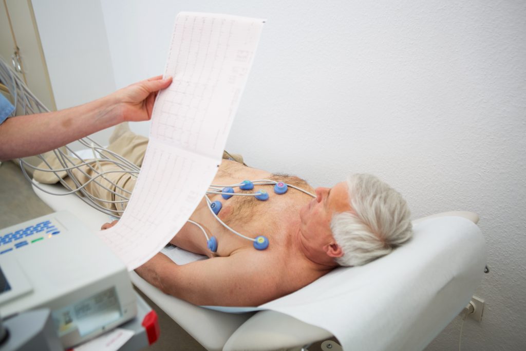
A standard resting ECG only records the heartbeat for a few seconds, so if atrial fibrillation is not present during this time prolonged monitoring may be required.
Heart monitoring
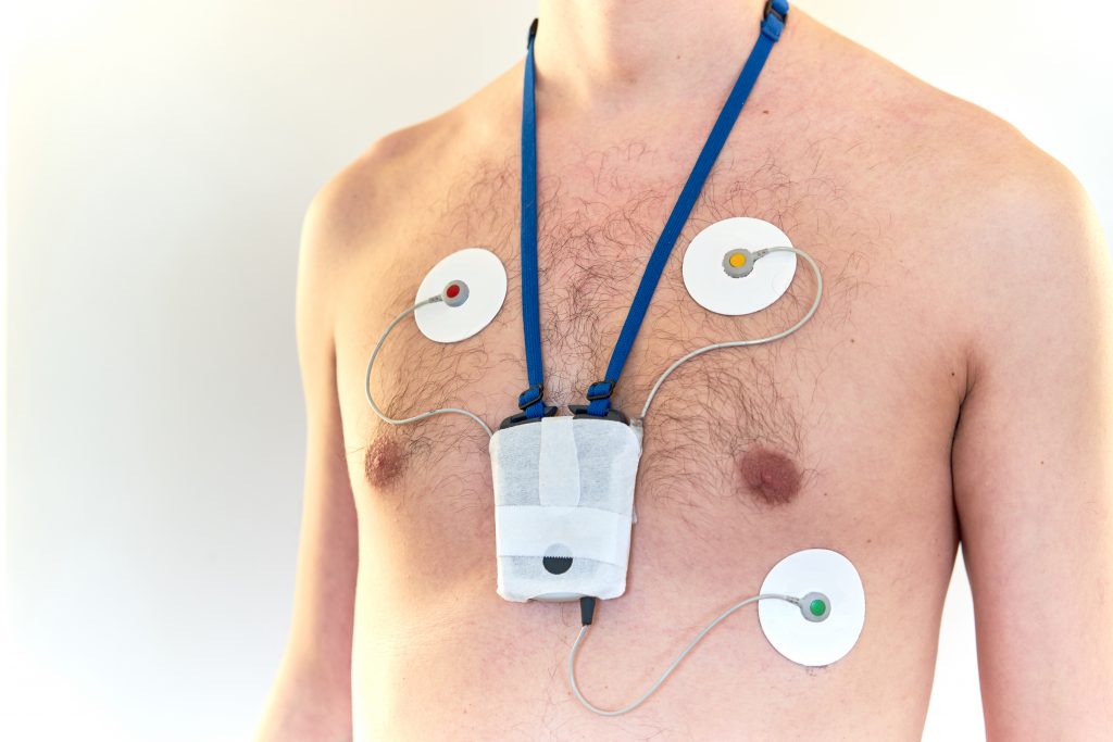
If your atrial fibrillation lasts just for a short period of time, it may be necessary to have a heart monitoring for 24 hours or longer (sometimes for 7 days or longer). This test, often called “Holter monitoring”, involves a small recording device attached by wires to 4 special stickers (electrodes) placed on your chest (see above). The device is easy to carry under clothes during your usual daily activities. A diary of your activities and any symptoms you experience while wearing the device can be useful. This test will provide your doctor with detailed information on your heart rhythm and correlation with your symptoms.
An alternative test is event monitoring. This monitor doesn’t continuously record, but will automatically record your ECG at certain times or when you manually trigger a recording, e.g. when you feel symptoms. These wearable event monitors can usually be used for up to 2 weeks.
If atrial fibrillation is still not detected but suspected an implantable cardiac monitor can be used for continuous heart rhythm monitoring. These monitors are positioned under the chest skin and can record changes in heart rhythm, usually for up to three years.
New technologies embedded in smartphones and smartwatches allow patients to have their own monitors (often called wearable devices) and record their heart rhythm on an ECG tracing. The reliability of these devices is being tested in clinical trials. In order to diagnose atrial fibrillation, these recordings should be evaluated by a physician.
Cardiac ultrasound
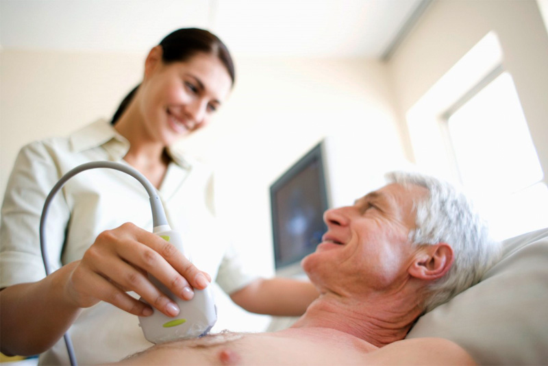
Echocardiogram
If you are diagnosed with atrial fibrillation, your doctor may want to examine your heart with an echocardiogram, often called an “echo” or an “ultrasound heart scan”. During this test a doctor or a cardiac technician will scan your chest using a handheld probe with some gel on it, in order to check the size and function of the four major chambers of the heart, the heart muscle and the valves.
An echocardiogram does not help your doctor detect atrial fibrillation, but it provides important information about the effect of atrial fibrillation on your heart and your prognosis.
Transoesophageal echocardiography
Sometimes your doctor will choose to investigate your heart with a transoesophageal echocardiogram, or ”TOE”. During this test a flexible tube is introduced through your mouth and positioned in your oesophagus (the passage leading from your mouth to your stomach). As the heart is positioned just in front of the oesophagus, TOE can give very detailed images of the most important parts of the heart. TOE is also the gold standard test to detect blood clots that may be forming inside the left atrium because of atrial fibrillation.
A TOE is often used before a cardioversion, if it is unclear when atrial fibrillation started and your doctor wants to be sure that no blood clots have already developed in your heart.
Other tests
Blood tests
Once atrial fibrillation is diagnosed you may also be asked to take some blood tests. Although the blood tests are not needed to make the diagnosis, they may help to explain why you have developed atrial fibrillation, for example due to thyroid gland problems or an imbalance of your body’s electrolytes. The blood tests will also help your doctor to choose the best treatment(s) for you.
Additional imaging of the heart
In some cases your doctor may want to perform a stress test in order to investigate what happens to your heart when it beats faster. Cardiac computer tomography or cardiac magnetic resonance imaging can also be used for detailed analysis of the structure and function of the heart. Whether further investigations are necessary depends on your condition and you should discuss it with your doctor.
FREQUENTLY ASKED QUESTIONS
What tests are used to diagnose AFib?
Common tests include an ECG (electrocardiogram) to record heart rhythm, Holter monitors for continuous monitoring, and echocardiograms to assess heart structure and function. Smart watches and wearables become increasingly popular in screening and detecting AFib as well.
Can blood tests detect AFib?
Blood tests cannot directly diagnose AFib but can identify contributing factors like thyroid issues, electrolyte imbalances, or inflammation.
Does AFib always show up on an ECG?
No, AFib may not appear on a single ECG if episodes are intermittent. Continuous monitoring with Holter monitors or event recorders is often required. Smartwatches euipted with an ECG function can also be used to detect AFib.
How does an echocardiogram help diagnose AFib?
An echocardiogram provides images of the heart to detect structural abnormalities, valve issues, or blood clots, which can contribute to or result from AFib. However, a echocardiogram is not used to diagnose AFib.
Can stress tests detect AFib?
Stress tests can help identify how your heart responds to exercise and whether AFib or other arrhythmias occur under stress.
Are at-home devices reliable for detecting AFib?
Many smartwatches and portable ECG devices can detect AFib, but they should not replace professional diagnosis. Discuss results with your doctor.
Related Pages
- Tests Used to Diagnose Atrial Fibrillation
- Restoring Normal Heart Rhythm in AFib
- Controlling Heart Rate in Atrial Fibrillation
- What Is Cardioversion for AFib?
- Atrial Fibrillation Ablation Explained
- Pacemakers & AV Node Ablation for AFib
- Preventing Stroke With Atrial Fibrillation
- Preventing Heart Failure in AFib
- Preventing AFib Recurrence