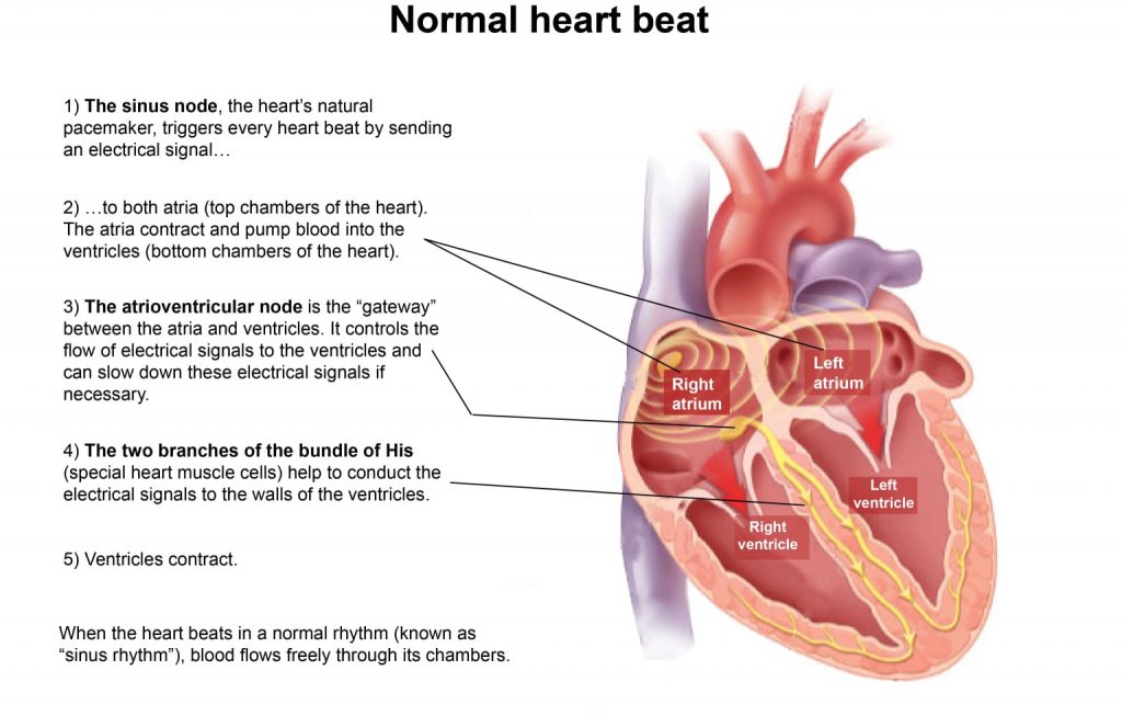
The heart consists of 4 chambers. The upper chambers are the right and left atrium, the lower chambers are the right and left ventricle.
The atria collect blood returning to the heart via the veins and fill the ventricles of the heart.
The heart rhythm starts in the sinus node, a group of specialised cells generating electrical activity.

The electrical wave spreads from the sinus node across the atria towards the heart’s main relay station the “atrio-ventricular node”. From this node the wave spreads towards the ventricles and the heart contracts to pump blood into the body.
The pulmonary veins are vessels transporting blood from the lungs towards the left atrium. Most patients have 4 veins and the junction of these veins with the left atrium may have abnormal electrical properties causing atrial fibrillation.

 Français
 Deutsch
 Español
 Italiano
 Nederlands
 Polski
 Русский
 Svenska
 Português
 Hrvatski
 Ελληνικα
 简体中文
简体中文 العربية
